
Robotic-Assisted Spine Surgery in New York City
Dr. Zeeshan Sardar, MD, MSc, F.R.C.S.C
Co-Chief of Spinal Deformity Surgery • NewYork-Presbyterian / Columbia University
Och Spine Hospital • New York, NY
Robotic-assisted spine surgery represents a fundamental advance in how complex spinal procedures are planned and executed. Dr. Sardar uses robotic navigation as a standard tool in his practice — not as an occasional option, but as the baseline technology for his most demanding cases. For patients undergoing scoliosis correction, spinal deformity reconstruction, or complex revision surgery, this means every screw is placed with a level of precision that was not achievable a generation ago.
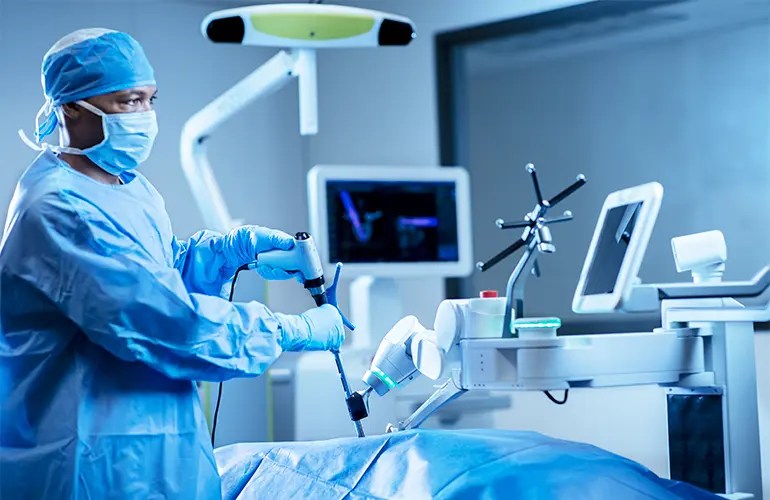
WHAT IS ROBOTIC SPINE SURGERY?
Robotic spine surgery does not mean a robot performs the operation. It means the surgeon uses a robotic guidance platform — combined with real-time imaging and computer navigation — to plan the surgery in three dimensions before making an incision, and then to execute that plan with exceptional accuracy during the procedure.
The core application is pedicle screw placement. Pedicle screws are the anchors that hold spinal instrumentation in place. They must be inserted through narrow bony corridors — the pedicles — on each vertebra. In deformity surgery, these corridors are often rotated, compressed, or structurally abnormal, making accurate placement technically demanding. Robotic guidance allows the surgeon to plan each screw’s trajectory on a CT-based 3D model of the patient’s spine before surgery, and then navigate each screw to its planned position with robotic-arm precision during the case.
HOW IT WORKS
Step 1: Pre-Operative Planning
Before surgery, a CT scan of the patient’s spine is uploaded to the robotic planning software. Dr. Sardar reviews the three-dimensional anatomy of each vertebra and plans the exact trajectory, depth, and diameter of every screw that will be placed. For a complex scoliosis case, this may involve planning multiple screws across multiple rotated vertebrae — all mapped out before the patient enters the operating room.
Step 2: Intraoperative Registration
Once the patient is positioned and the spine is exposed, the robotic system is registered to the patient’s anatomy using intraoperative imaging. This aligns the pre-operative plan with the actual spine on the table, accounting for any positional changes. Registration is verified before any instrumentation begins.
Step 3: Guided Screw Placement
The robotic arm moves to the planned position for each screw and holds the drill guide in the exact orientation specified in the pre-operative plan. Dr. Sardar then drills and places the screw through the robot-guided cannula. The surgeon’s hands perform the work — the robot ensures the trajectory is precisely maintained throughout.
Step 4: Real-Time Verification
After instrumentation is complete, intraoperative CT imaging confirms the position of every screw before the deformity correction is performed. Any screw that does not meet positional standards is revised immediately — before the patient leaves the operating room. This level of verification was not available with conventional fluoroscopic technique.
WHY IT MATTERS FOR PATIENTS
Greater Precision in Complex Anatomy
In spinal deformity surgery, the vertebrae are rotated and the pedicles are often narrow or distorted. Freehand screw placement in this anatomy — even by experienced surgeons — carries a meaningful rate of screw malposition. Published literature consistently demonstrates that robotic guidance reduces pedicle screw malposition rates compared to conventional freehand technique, with particularly significant gains in the most challenging deformity cases where accuracy matters most.
Reduced Radiation Exposure
Traditional spine surgery relies on continuous intraoperative fluoroscopy (live X-ray imaging) to guide screw placement. Robotic navigation significantly reduces the need for repeated fluoroscopic shots during the case. The shift to a single intraoperative CT scan for verification — rather than repeated fluoroscopy throughout — meaningfully reduces cumulative radiation exposure for both the patient and the surgical team.
Confidence in the Most Difficult Cases
The value of robotic guidance is greatest in the cases where accuracy is hardest to achieve: revision surgery through scar tissue, severely rotated deformity vertebrae, osteoporotic bone with poor tactile feedback, and the upper thoracic spine where the pedicles are narrowest. These are precisely the cases that make up a large part of Dr. Sardar’s practice. Using robotic guidance as a standard tool — not just for straightforward cases — means patients with the most complex anatomy benefit the most.
A System of Overlapping Safeguards
Robotic guidance does not stand alone. Dr. Sardar combines it with intraoperative neuromonitoring (IONM) — continuous real-time monitoring of spinal cord and nerve function throughout the procedure — and intraoperative 3D CT imaging for final verification. These three technologies work together as a system of overlapping safeguards, each catching what the others might miss.
CONDITIONS TREATED WITH ROBOTIC GUIDANCE
Dr. Sardar uses robotic-assisted navigation across the full spectrum of his surgical practice, including:
- Adolescent idiopathic scoliosis (AIS) — posterior spinal fusion with robotic pedicle screw placement across rotated deformity vertebrae
- Adult scoliosis and spinal deformity — long-segment fusions requiring consistent accuracy across many levels
- Kyphosis correction — including cases requiring osteotomy, where screw purchase above and below the correction site is critical
- Revision spine surgery — where prior surgery, scar tissue, and altered anatomy make freehand technique particularly challenging
- Harrington rod revision — complex long-segment reconstruction in patients with distorted anatomy from prior instrumentation
- Cervical and lumbar fusion — for degenerative conditions, instability, and stenosis requiring stabilization
- Minimally invasive spine surgery — where limited visualization makes robotic navigation particularly valuable for maintaining accuracy through small incisions
FREQUENTLY ASKED QUESTIONS
Does the robot perform the surgery?
No. The robot does not make decisions, perform cuts, or operate independently. It is a guidance tool. Dr. Sardar plans, executes, and controls every aspect of the surgery. The robotic arm holds the drill guide in the planned position while he places each screw — the precision is his to direct, with the robot ensuring the trajectory is maintained. The surgical judgment, technique, and decision-making are entirely the surgeon’s.
Is robotic spine surgery right for me?
Robotic guidance is most valuable in complex cases involving deformity, revision surgery, or anatomy that makes freehand placement particularly demanding. Dr. Sardar will discuss whether robotic assistance is planned for your specific procedure at your pre-operative visit. For most of his spinal deformity and revision cases, it is part of the standard approach.
Does robotic surgery mean a smaller incision?
Not necessarily. Robotic guidance and minimally invasive techniques are separate — though they are often used together. For complex deformity corrections requiring osteotomy and long-segment fusion, a larger exposure is typically needed regardless of whether robotic guidance is used. The benefit of robotics in these cases is precision, not incision size. For selected patients undergoing less complex procedures, robotic guidance and minimally invasive approaches can be combined to reduce tissue disruption and recovery time.
How do I know if my surgeon uses robotics?
Ask directly. The right questions are: Do you use robotic navigation for this procedure? Do you verify screw position with intraoperative CT? How many robotic cases have you performed? A surgeon who uses these technologies routinely will answer these questions clearly and specifically.
ROBOTIC SURGERY AT NEWYORK-PRESBYTERIAN
The Och Spine Hospital at NewYork-Presbyterian is one of the most technologically advanced spine centers in the United States. The combination of robotic navigation platforms, intraoperative CT imaging, and dedicated neuromonitoring teams makes it one of the few environments in the country where the full technology stack for complex deformity surgery is consistently available and reliably integrated into daily practice.
For patients traveling from out of state or internationally, this infrastructure — combined with Dr. Sardar’s experience and the institution’s depth of subspecialty support — is part of why NewYork-Presbyterian is the destination they choose for their most complex spine cases.
This page is for educational purposes only and does not constitute individualized medical advice. Please consult a qualified spine specialist to discuss your specific condition and treatment options.
REQUEST A CONSULTATION
To schedule a consultation with Dr. Sardar, call 212-932-5187 or use the contact form below.
